Criteria assessed to evaluate the anatomical consistency by positional relationship
This group of criteria is based on positional relationship analysis to assess craniofacial anatomical consistency. In other words, the objective is to evaluate the relative position of a skull region against a facial region and determine the degree of anatomical consistency of the relative position of both regions. For example, the lateral corner of the eye should be within the lateral margin of the orbit in lateral and oblique view.
-
Criteria assessed in Frontal View:
- The prosthion lies posterior to the anterior edge of the upper lip. The occlusal and the lip closure line are consistent.
- The piriform aperture width and height lies within the borders of the nose.
- The stomion lies at the central incisors (incisal margin of the upper incisors).
- The medial margin of the orbits aligns and superimposes with the endocanthion.
- The Whitnall's tubercle aligns with the exocanthion on the horizontal plane and vertically the exocanthion lies medial to the tubercle. The orbital width is consistent with the eye-slit width.
- The cheilion lies between the canine and the first premolar (at the occlusal line).
- The eyebrow generally follows the upper edge of the orbit over the medial two-thirds. At lateral superior one-third of the orbit the eyebrow continues horizontally as the orbital rims begins to curve inferiorly.
- The external auditory meatus opening lies medially to the tragus of the ear (place a projecting marker at the ear canal to assess this criterion more easily).
- Gonial flare in the skull and the postero-lateral jaw prominence in the face.
-
Criteria assessed in Lateral/Oblique View:
- The lateral angle of the eye lies within the lateral wall of the orbit.
- The lateral orbital margin at the Whitnall's tubercle matches or approximates the position of the exocanthion.
- The stomion lies at the central incisors (at the occlusal line).
- The lateral margin of the piriform aperture matches or approximates the alare.
- The prosthion lies posterior to the anterior edge of the upper lip. The occlusal and the lip closure line are consistent.
- The porion aligns just posterior to the tragus, slightly inferior to the crus of the helix.
- The lower margin of piriform aperture matches the subnasale.
- El cheilion se encuentra entre el canino y el primer premolar (en la línea oclusal).
- The anterior nasal spine lies posterior to the base of the nose near the most posterior portion of the lateral septal cartilage.
- Gonial flare in the skull and the postero-lateral jaw angle outline in the face.
Criteria assessed in Frontal View
This group includes a series of anatomical criteria that can be evaluated in a frontal view photograph.
- The prosthion lies posterior to the anterior edge of the upper lip. The occlusal and the lip closure line are consistent. This criterion has a good trade-off between subjectivity and discriminative power according to Ibáñez y col. (2016).
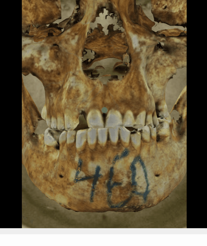 Figure 1. Example of a positive match in which the prosthion position is evaluated with the transparency tool, showing that the prosthion lies posterior to the anterior edge of the upper lip (cupid’s bow) in a consistent way. The transparency tool has been used to show a gradient of opacity over the teeth showing that the prosthion in the skull is positioned in the anterior edge of the upper lip in the face.
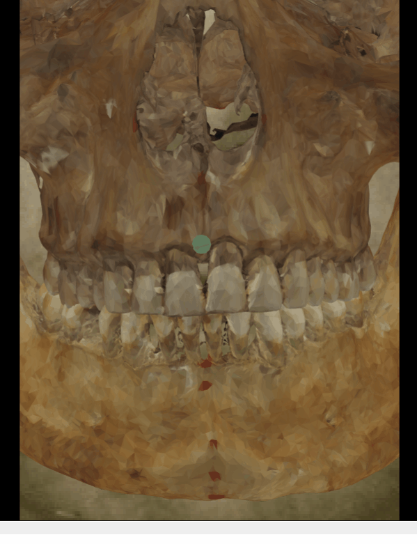 Figure 2. Example of a positive match in which the occlusal line is evaluated with the transparency and wipe tools, showing that the occlusal and the lip closure line are consistent. The transparency tool has been used in combination with the wipe tool to show a gradient from the right corner (anatomical) of the mouth to the left corner monitoring the occlusal line over the lip closure line.
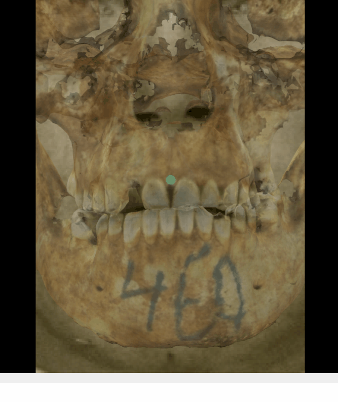 Figure 3. Example of a negative match in which the prosthion position is evaluated with the transparency tool, showing that the prosthion does not lie posterior to the anterior edge of the upper lip (cupid’s bow) in a consistent way. The transparency tool has been used to show a gradient of opacity over the teeth showing that the prosthion in the skull is positioned some mms above the anterior edge of the upper lip in the face.
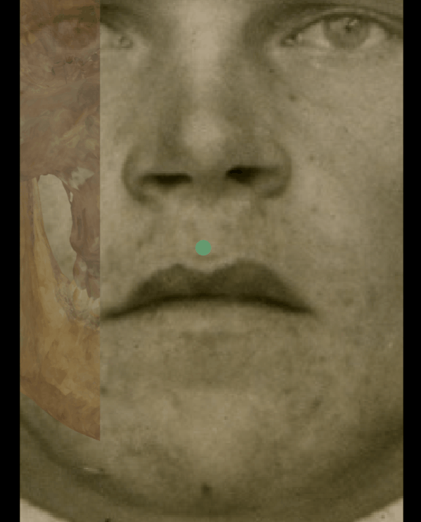 Figure 4. Example of a negative match in which the occlusal line is evaluated with the transparency and wipe tools, showing that the occlusal and the lip closure line are inconsistent. The transparency tool has been used in combination with the wipe tool to show a gradient from the right corner (anatomical) of the mouth to the left corner monitoring the occlusal line over the lip closure line.
- The piriform aperture width and height lies within the borders of the nose.
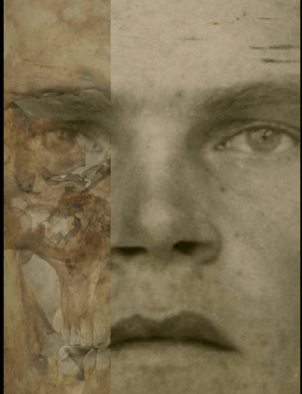 Figure 5. Example of a positive match in which the piriform aperture width and height is visually evaluated with Skeleton·ID by means of the transparency and wipe tools, showing that the piriform aperture width and height lies within the borders of the nose in a consistent way. The transparency tool has been used in combination with the wipe tool to show a gradient from the right to the left (anatomical) alare showing the position of the piriform aperture in relation to the nose in the face.
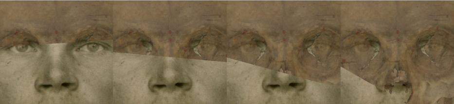 Figure 6. Example of a negative match in which the piriform aperture width and height is visually evaluated with Skeleton·ID by means of the transparency and wipe tools, showing that the piriform aperture width and height does not lie within the borders of the nose. The transparency tool has been used in combination with the wipe tool to show a gradient showing the position of the piriform aperture in relation to the nose in the face.
- The stomion lies at the central incisors (incisal margin of the upper incisors). This criterion has a good trade-off between subjectivity and discriminative power according to Ibáñez y col. (2016).
 Figure 7. Example of a positive match in which the stomion is visually evaluated with Skeleton·ID by means of the transparency tool, showing that the stomion in the face lies at the central incisors (incision landmark) in a consistent way. The transparency tool has been used to show a gradient of opacity over the teeth showing the position of stomion in relation to the central incisors.
 Figure 8. Example of a negative match in which the stomion is visually evaluated with Skeleton·ID by means of the transparency tool, showing that the stomion in the face does not lie at the position of the central incisors (incision landmark) in a consistent way. The transparency tool has been used to show a gradient of opacity over the teeth showing the position of stomion in relation to the central incisors. In this case the incisor is located some mms below of the position lip closure line.
- The medial margin of the orbits aligns and superimpose with the endocanthion.
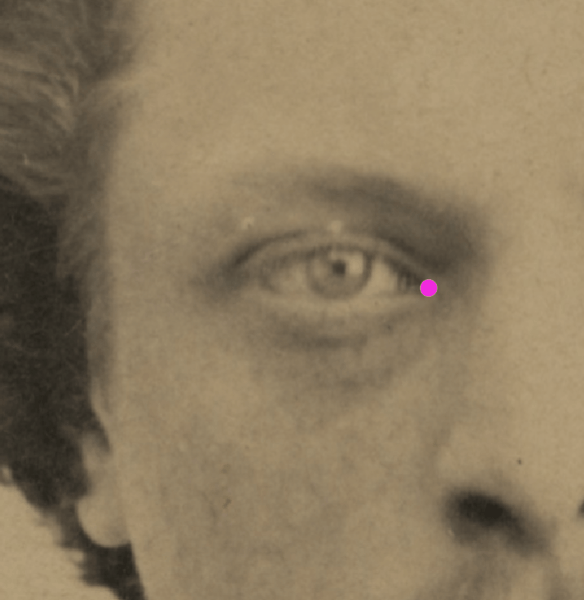 Figure 9. Example of a positive match in which the endocanthion is visually evaluated with Skeleton·ID by means of the transparency tool, showing that the right endocanthion in the face is aligned and superimposed over the medial margin of the orbit. The transparency tool has been used to show a gradient of opacity of the orbit.
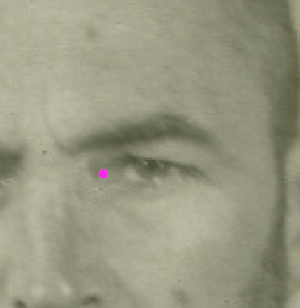 Figure 10. Example of a negative match in which the endocanthion is visually evaluated with Skeleton·ID by means of the transparency tool, showing that the right endocanthion in the face is not aligned with the medial margin of the orbit. The transparency tool has been used to show a gradient of opacity of the orbit.
- The Whitnall´s tubercle aligns with the exocanthion on the horizontal plane and vertically the exocanthion lies medial to the tubercle. The orbital width is consistent with the eye-slit width. This criterion has a good trade-off between subjectivity and discriminative power according to Ibáñez y col. (2016).
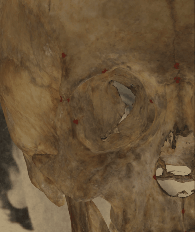 Figure 11. Example of a positive match in which the exocanthion is visually evaluated with Skeleton·ID by means of the transparency tool, showing that the right exocanthion in the face is aligned with the Whitnall´s tubercle on the horizontal plane. The orbital width is consistent with the eye-slit width. The transparency tool has been used to show a gradient of opacity of the orbit.
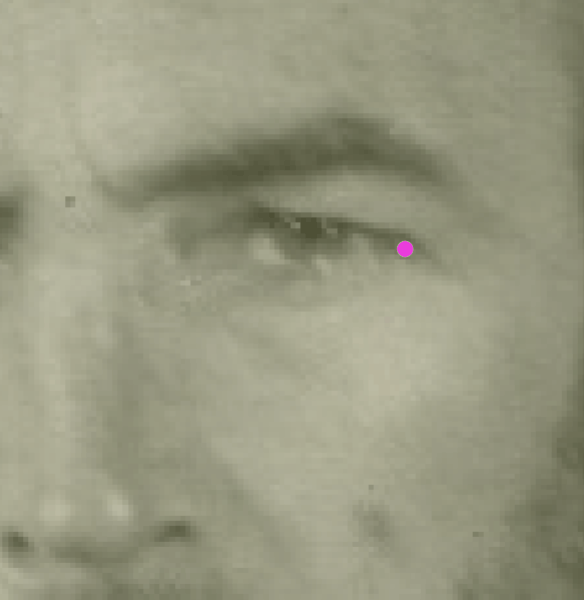 Figure 12. Example of a negative match in which the exocanthion is visually evaluated with Skeleton·ID by means of the transparency tool, showing that the left exocanthion in the face is not aligned with the Whitnall´s tubercle on the horizontal plane. In this case the exocanthion falls inside the orbit. The transparency tool has been used to show a gradient of opacity of the orbit.
- The cheilion lies between the canine and the first premolar (at the occlusal line).
 Figure 13. Corners on radiating lines from first premolar-canine junction (Krogman e Iscan, 1986).
- The eyebrow generally follows the upper edge of the orbit over the medial two-thirds. At lateral superior one-third of the orbit the eyebrow continues horizontally as the orbital rims begins to curve inferiorly. This criterion should be considered with caution due to the low discriminative power and subjectivity in frontal view according to Ibáñez y col. (2016).
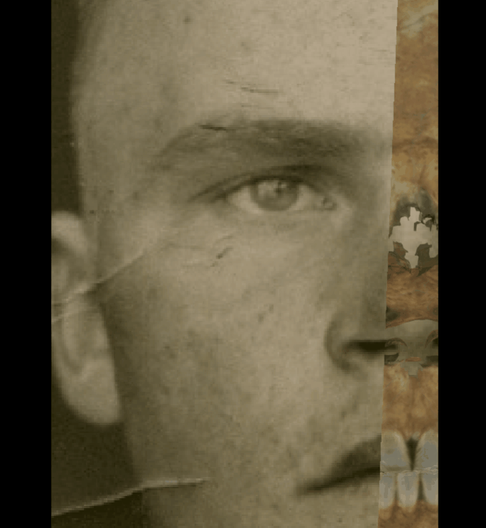 Figure 14. Example of a positive match in which the right eyebrow is visually evaluated with Skeleton·ID by means of the transparency and wipe tools, showing that the right eyebrow follows the upper edge of the orbit and at the lateral superior one-third of the orbit it continues horizontally while the orbital ring descends in in a consistent way. The transparency tool has been used in combination with the wipe tool to show a gradient from the medial side of the orbital rim to the lateral side monitoring the eyebrow position.
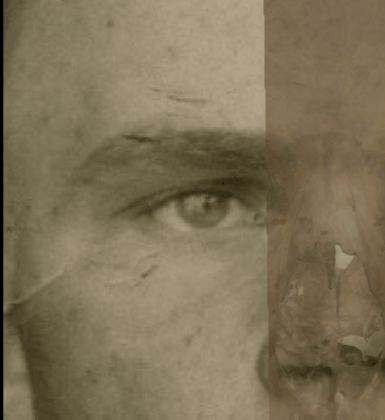 Figure 15. Example of a negative match in which the right eyebrow is visually evaluated with Skeleton·ID by means of the transparency and wipe tools, showing that the right eyebrow follows the upper edge of the orbit and at the lateral superior one-third of the orbit it does not continues horizontally as it is covered by the orbital ring in an inconsistent way. The transparency tool has been used in combination with the wipe tool to show a gradient from the medial side of the orbital rim to the lateral side monitoring the eyebrow position.
- The external auditory meatus opening lies medial to the tragus of the ear (place a projecting marker at the ear canal to asses this criterion more easily).
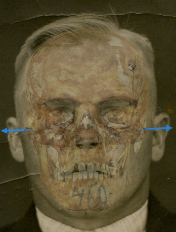 Figure 16. Example of a positive match in which the position of the external auditory meatus is visually evaluated with Skeleton·ID by means of the transparency tool, showing that the external auditory meatus opening lies medial to the tragus of the ear in a consistent way. The direction of the vectors of both porion has been used as a projecting markers of the ear canal to asses this criterion more easily.
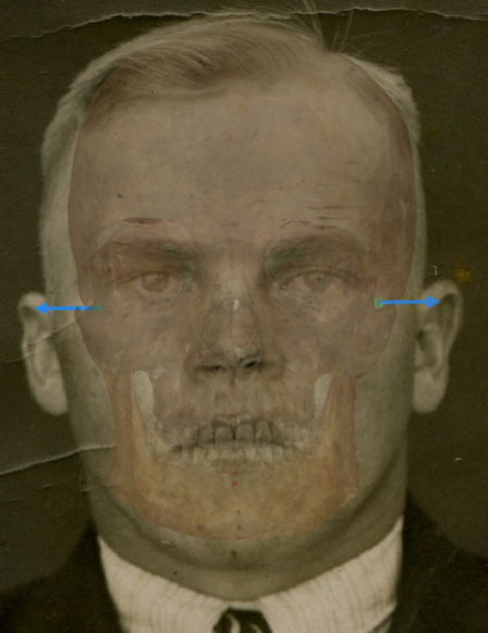 Figure 17. Example of a negative match in which the position of the external auditory meatus is visually evaluated with Skeleton·ID by means of the transparency tool, showing that the external auditory meatus opening does not lie medial to the tragus of the ear in a consistent way. The direction of the vectors of both porion has been used as a projecting markers of the ear canal to asses this criterion more easily. In this case we can see how both auditory meati are positioned at the helix level (and not at the tragus level) in both ears, showing a positional inconsistency between both structures.
- Gonial flare in the skull and the postero-lateral jaw prominence in the face. This criterion has a good trade-off between subjectivity and discriminative power according to Ibáñez y col. (2016).
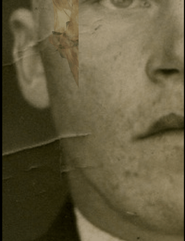 Figure 18. Example of a positive match in which the position of the gonial flare is visually evaluated with Skeleton·ID by means of the transparency and wipe tools, showing that the gonial flare in the skull and the postero-lateral jaw prominence in the face are consistent. The transparency tool has been used in combination with the wipe tool to show a gradient of the right jaw.
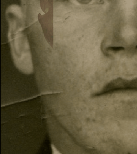 Figure 19. Example of a negative match in which the position of the gonial flare is visually evaluated with Skeleton·ID by means of the transparency and wipe tools, showing that the gonial flare in the skull and the postero-lateral jaw prominence in the face are inconsistent. The transparency tool has been used in combination with the wipe tool to show a gradient of the right jaw. In this case the gonial flare in the skull is not located at the same position of the postero-lateral prominence of the jaw in the face.
Criteria assessed in Lateral/Oblique View
This group includes a series of anatomical criteria that can be evaluated in a lateral and / or oblique view photograph.
- The lateral angle of the eye lies within the lateral wall of the orbit.
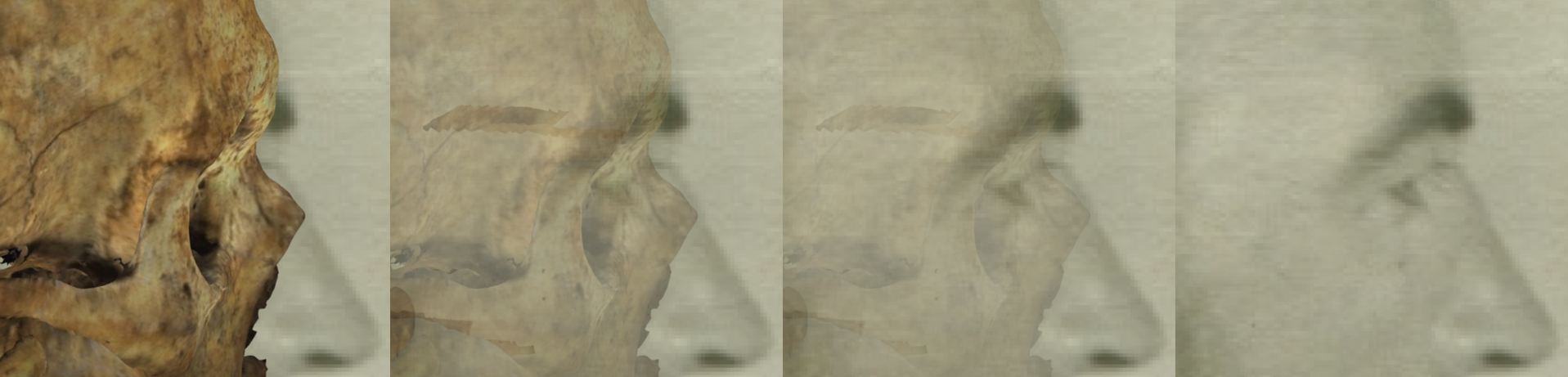 Figure 20. Example of a positive match in which the position of the lateral angle of the eye is visually evaluated with Skeleton·ID by means of the transparency tool, showing that the lateral angle of the eye lies within the lateral wall of the orbit in a consistent way. The transparency tool has been used to show a gradient of opacity of the orbital rim.
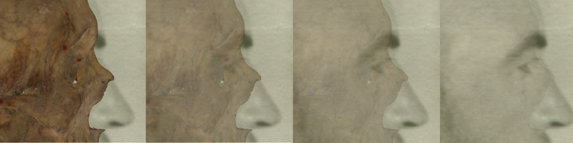 Figure 21. Example of a negative match in which the position of the lateral angle of the eye is visually evaluated with Skeleton·ID by means of the transparency tool, showing that the lateral angle of the eye does not fit with the lateral wall of the orbit. The transparency tool has been used to show a gradient of opacity of the orbital rim.
- The lateral orbital margin at the Whitnall´s tubercle matches or approximates the position of the exocanthion.
 Figure 22. Example of a positive match in which the position of the right exocanthion is visually evaluated with Skeleton·ID by means of the transparency tool, showing that the position of the rigth exocanthion approximates the right lateral margin of the orbit in a consistent way. The transparency tool has been used to show a gradient of opacity of the orbital rim.
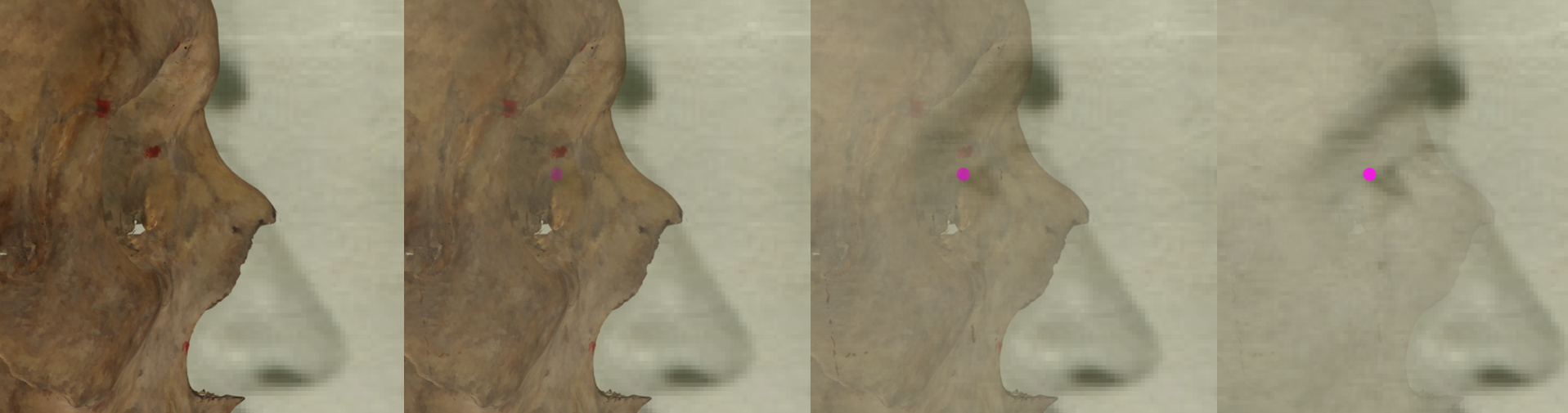 Figure 23. Example of a negative match in which the position of the right exocanthion is visually evaluated with Skeleton·ID by means of the transparency tool, showing that the position of the rigth exocanthion approximates the right lateral margin of the orbit in a consistent way. The transparency tool has been used to show a gradient of opacity of the orbital rim.
- The stomion lies at the central incisors (at the occlusal line).
 Figure 24. Example of a positive match in which the stomion is visually evaluated with Skeleton·ID by means of the transparency tool, showing that the stomion in the face lies at the central incisors (incision landmark) in a consistent way. The transparency tool has been used to show a gradient of opacity over the teeth showing the position of stomion in relation to the central incisors.
 Figure 25. Example of a negative match in which the stomion is visually evaluated with Skeleton·ID by means of the transparency tool, showing that the stomion in the face does not lie at the central incisors in a consistent way. In this case the stomion lies at the left central incisive. The transparency tool has been used to show a gradient of opacity over the teeth showing the position of stomion in relation to the central incisors.
- The lateral margin of the piriform aperture matches or approximates the alare.
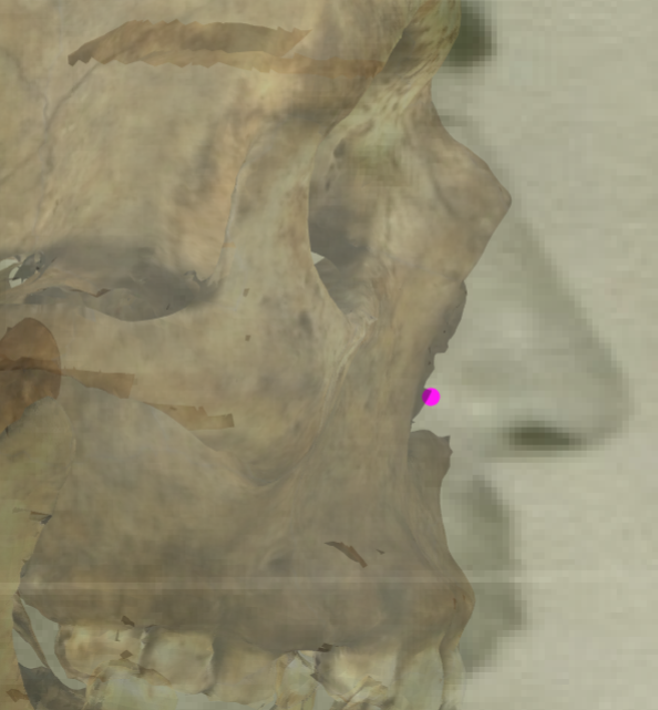 Figure 26. Example of a positive match in which the position of the right alare is visually evaluated with Skeleton·ID by means of the transparency tool, showing that the right lateral margin of the piriform aperture approximates the right alare in a consistent way.
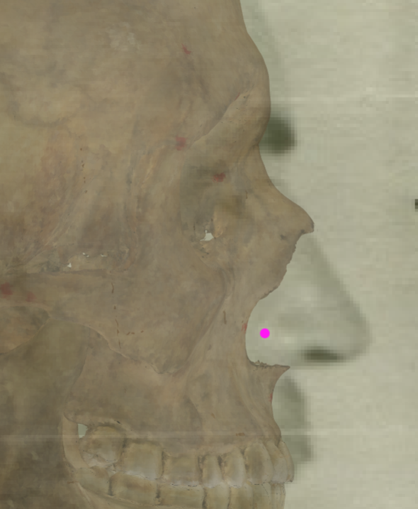 Figure 27. Example of a negative match in which the position of the right alare is visually evaluated with Skeleton·ID by means of the transparency tool, showing that the right lateral margin of the piriform aperture does not match with the right alare.
- The prosthion lies posterior to the anterior edge of the upper lip. The occlusal and the lip closure line are consistent.
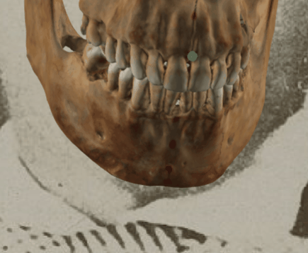 Figure 28. Example of a positive match in which the position of the prosthion is visually evaluated with Skeleton·ID by means of the transparency tool, showing that the prosthion lies posterior to the anterior edge of the upper lip in a consistent way. The transparency tool has been used to show a gradient of opacity of the teeth over the mouth.
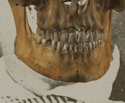 Figure 29. Example of a negative match in which the position of the prosthion is visually evaluated with Skeleton·ID by means of the transparency tool, showing that the prosthion does not lie posterior to the anterior edge of the upper lip. The transparency tool has been used to show a gradient of opacity of the teeth over the mouth.
- The porion aligns just posterior to the tragus, slightly inferior to the crus of the helix. This criteron has a highest discriminatory power but also high variance according to Ibáñez y col. (2016).
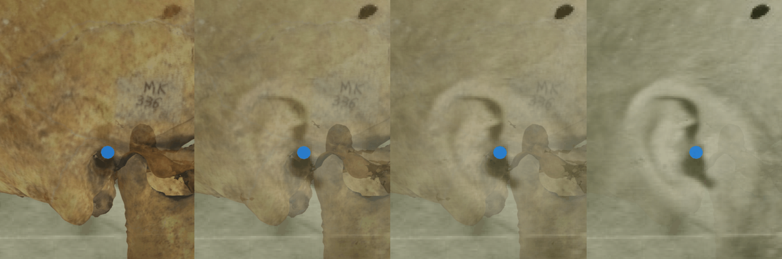 Figure 30. Example of a positive match in which the position of the right porion is visually evaluated with Skeleton·ID by means of the transparency tool, showing that the right porion aligns posterior to the tragus, slightly inferior to the crus of the helix in a consistent way. The transparency tool has been used to show a gradient of opacity of the auditive meatus showing the position of the porion over the ear.
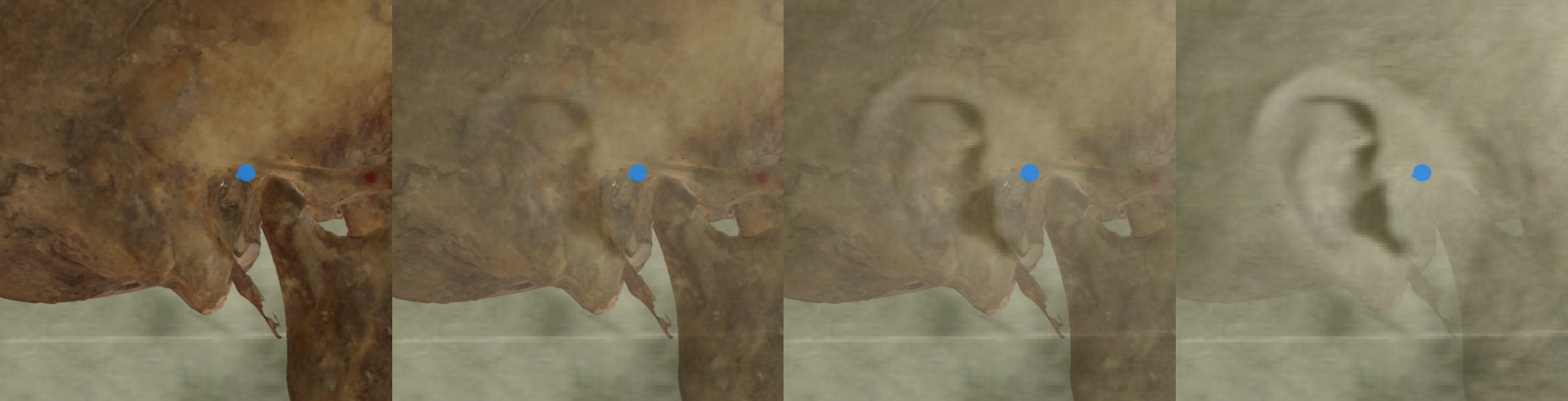 Figure 31. Example of a negative match in which the position of the right porion is visually evaluated with Skeleton·ID by means of the transparency tool, showing that the right porion does not align posterior to the tragus, in this case is located in an upper right area. The transparency tool has been used to show a gradient of opacity of the auditive meatus showing the position of the porion over the ear.
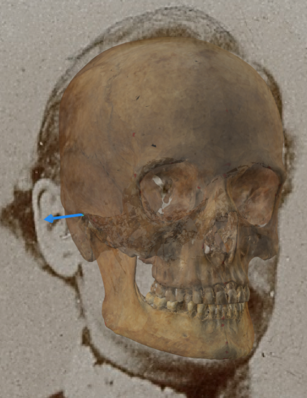 Figure 32. Example of a positive match in which the position of the right porion is visually evaluated with Skeleton·ID by means of the transparency tool, showing that the right porion aligns posterior to the tragus, slightly inferior to the crus of the helix in a consistent way. The transparency tool has been used to show a gradient of opacity of the auditive meatus showing the position of the porion over the ear.
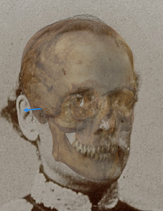 Figure 33. Example of a negative match in which the position of the right porion is visually evaluated with Skeleton·ID by means of the transparency tool, showing that the right porion does not align posterior to the tragus, and it is located over the crus of the helix in an inconsistent way. The transparency tool has been used to show a gradient of opacity of the auditive meatus showing the position of the porion over the ear.
- The lower margin of piriform aperture matches the subnasale. This criterion should be considered with caution due to the low discriminative power and subjectivity in oblique view according to Ibáñez y col. (2016).
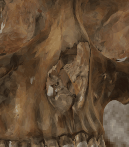 Figure 34. Example of a positive match in which the subnasale position is visually evaluated with Skeleton·ID by means of the transparency tool, showing that the subnasale matches the lower margin of the piriform aperture in a consistent way. The transparency tool has been used to show a gradient of opacity of the piriform aperture.
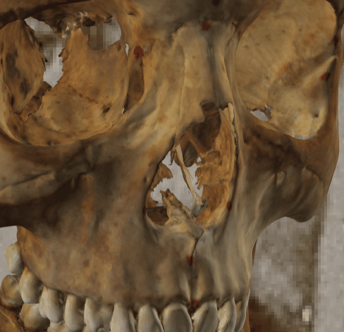 Figure 35. Example of a negative match in which the subnasale position is visually evaluated with Skeleton·ID by means of the transparency tool, showing that the subnasale does not matche the lower margin of the piriform aperture. The transparency tool has been used to show a gradient of opacity of the piriform aperture.
- The anterior nasal spine lies posterior to the base of the nose near the most posterior portion of the lateral septal cartilage.
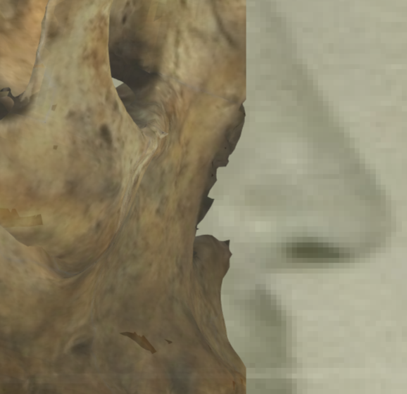 Figure 36. Example of a positive match in which the position of the anterior nasal spine is visually evaluated with Skeleton·ID by means of the transparency tool, showing that the anterior nasal spine lies posterior to the base of the nose, near the most posterior portion of the septal cartilage, in a consistent way.
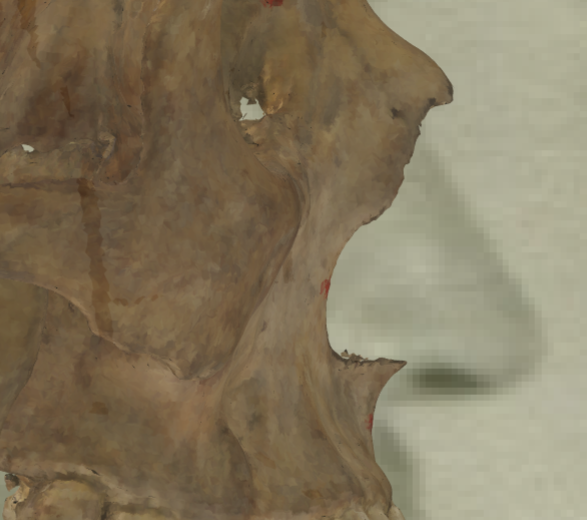 Figure 37. Example of a negative match in which the position of the anterior nasal spine is visually evaluated with Skeleton·ID by means of the transparency tool, showing that the anterior nasal spine lies over the base of the nose.
- Gonial flare in the skull and the postero-lateral jaw angle outline in the face.
 Figure 38. Example of a positive match in which the position of the gonial flare is visually evaluated with Skeleton·ID by means of the wipe tool, showing that the gonial flare in the skull and the postero-lateral jaw prominence in the face are consistent. The wipe tool has been used to show a gradient of the right jaw.
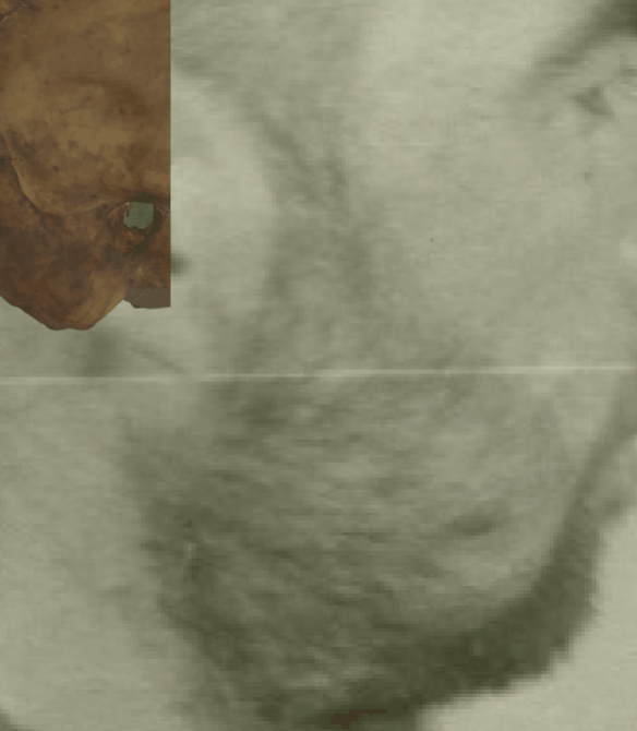 Figure 39. Example of a negative match in which the position of the gonial flare is visually evaluated with Skeleton·ID by means of the transparency and wipe tools, showing that the gonial flare in the skull and the postero-lateral jaw prominence in the face are inconsistent. The wipe tool has been used to show a gradient of the right jaw.
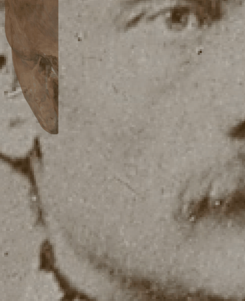 Figure 40. Example of a positive match in which the position of the gonial flare is visually evaluated with Skeleton·ID by means of the wipe tool, showing that the gonial flare in the skull and the postero-lateral jaw prominence in the face are consistent. The wipe tool has been used to show a gradient of the right jaw.
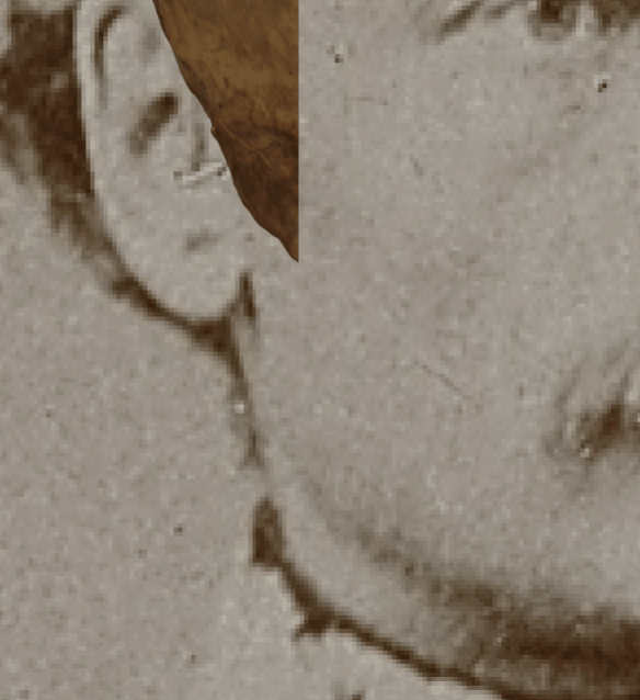 Figure 41. Example of a negative match in which the position of the gonial flare is visually evaluated with Skeleton·ID by means of the wipe tool, showing that the gonial flare in the skull and the postero-lateral jaw prominence in the face are inconsistent. In this case the morphology and the position of the gonial flare are inconsistent with the jaw prominence in the face. The transparency tool has been used in combination with the wipe tool to show a gradient of the right jaw.